Multi-Orthogonal Analytics are Essential for viral vector characterization because the inherent complexity of capsid viral proteins and genomic integrity cannot be captured by a single test.
Critical Quality Attributes (CQAs) must include viral potency, identity, empty/full capsid ratios, and process residuals to meet stringent regulatory specifications for product release.
Digital Droplet PCR (ddPCR) has become the industry standard for absolute quantitation of vector genome titers and residual DNA because it provides higher precision and sensitivity without requiring external standards.
Capillary Electrophoresis (CE-LIF/CE-SDS) is replacing traditional methods like SDS-PAGE and agarose gel electrophoresis due to its superior speed, automation, and resolution in analyzing pDNA isoforms and capsid purity.
Potency Assays remain a significant analytical challenge because they must measure biological activity in specific cell types, which varies depending on the clinical indication and determines the final patient dosing.
1. Analytics for AAV Production
1.1. As with any biologic drug substance and drug product, raw material analysis, in-process monitoring, and product-release testing are essential to ensuring the quality and safety of AAV vectors used for gene therapy.
1.2. The quality and purity of the plasmid DNA, culture media, transfection agents, and other raw materials must be confirmed. All starting materials and components must be qualified and suitable for the intended purpose.
1.3. Analytics are required for in-process monitoring during cell culture, harvest, and each downstream unit operation during AAV production. Product-release testing is necessary to confirm that viral vectors meet specifications required for use in the genetic modification of cells related to specific vector properties, impurity levels (e.g., HCPs, host cell DNA), and contaminants, such as mycoplasma and adventitious agents. Critical quality attributes (CQAs) for viral vectors include viral potency, identity, quantity, process residuals, aggregation, empty capsids, protein content, and product safety.
1.4. AAV capsid viral proteins are crucial to the performance of these viral vectors as gene therapies. They are involved in receptor binding during cell entry, intracellular trafficking, and genome release and thus determine treatment efficacy. Consequently, full characterization of the capsid proteins and their purity is essential.
1.5. To achieve this goal, the purity and structure of the pDNA used to produce the capsids must be confirmed. Similarly, the size, peptide sequence, and posttranslational modifications (PTMs) of the capsid, as well as its purity with respect to HCPs, host cell DNA and RNA, and partial and empty capsids must be evaluated. Potency assays — traditionally cell-based — must also be determined for the final vector products. Different needs for in-process and product-release analyses and a lack of specific regulatory requirements further complicate the analytical landscape.
1.6. This complexity necessitates the use of multiple orthogonal methods to fully understand the physicochemical properties and quality of viral vectors.[1]
1.7. Capillary electrophoresis (CE) with laser-induced fluorescence detection (CE-LIF) is a rapid, sensitive, reproducible, and automated method for the quantitative analysis of pDNA isoforms that can provide separation and identification of supercoiled, linearized, and open circular plasmid isoforms for 5-kb and 7-kb plasmids with baseline resolution in less than 15 minutes.[2][3]New solutions for automated Sanger genome sequencing also enable rapid sequencing of recombinant DNA in plasmid vectors. One example is the SCIEX GenomeLab GeXP Genetic Analysis System, which can be used for routine as well as more complex analyses.[4]
1.8. Quadrupole time-of-flight mass spectrometry (Q-TOF MS) enables rapid characterization of viral vector proteins. Using proprietary SWATH® Acquisition technology from SCIEX combined with the company’s BioPharmaView™ software, peptide mapping can be achieved with a single sample injection, with high-resolution mass spectroscopy (MS) and MS/MS data obtained for all low-abundance peptides and PTMs often missed when using information-dependent peptide map workflows.[5]
1.9. HCPs and other contaminants can also be readily identified and quantified using SWATH-based liquid chromatography with tandem mass spectrometry (LC-MS/MS).[6] Capillary electrophoresis–sodium dodecyl sulfate (CE-SDS) is a supplementary method analogous to traditional sodium dodecyl sulfate–polyacrylamide gel electrophoresis (SDS-PAGE) techniques that allows for automated sample preparation and protein separation, reducing the labor and time required for this analysis while affording good sensitivity for the lower concentrations of viral proteins found in AAV samples, with higher resolution and better reproducibility.[7] More conventional methods include enzyme-linked immunosorbent assay (ELISA) and quantitative PCR; transmission electron microscopy (TEM) has also been applied for this purpose.[8]
1.10. Meanwhile, denaturing agarose gel electrophoresis and Southern blot methods are being replaced with more rapid and automated CE-LIF methods for assessing the quality and length/size of encapsulated AAV genomes and detecting intact and partial AAV genomes and smaller impurities with high resolution.[9] Next-generation sequencing (NGS) techniques are another option for identifying nucleic acid impurities, often required to sequence plasmids containing ITR sequences since Sanger sequencing sometimes does not work well on ITR regions.[1] A further rapid, effective, and reliable alternative to spectrophotometry, electron microscopy, IEX chromatography, analytical ultracentrifugation, and other methods for empty/partial/full capsid analysis is capillary isoelectric focusing (cIEF).[10]
1.11. AAV vector products must also be assessed for potency, typically using cell-based assays; physical titer, using methods such as digital droplet polymerase chain reaction (ddPCR), the optical density (A260/280) assay, and high-performance liquid chromatography (HPLC); and infectious or functional titer [expressed as plaque-forming units per mL (PFU/mL) or infectious units per mL (IFU/mL) for AAV vectors], determined most often using the viral plaque assay, but also the endpoint dilution assay (TCID50) and the immunofluorescence assay (IFA).[8]
1.12. Potency is difficult to assay, because different methods provide different results due to the need to measure biological activity in any number of specific cell types depending upon the indication. However, it is a crucial assay because it determines dosing. Gene therapies based on AAVs are most commonly dosed based on the vector genomes, but results can depend on the method used to determine the genome concentration.
1.13. Assays for dose determination are product-dependent and must be optimized for the specific AAV vector and qualified before initiating clinical studies, including demonstration of assay linearity, accuracy, precision, range, sensitivity, and specificity.
1.14. Safety testing is required for all viral vectors, just as it is for all biologics, including sterility, endotoxin, mycoplasma, adventitious virus, and replication-competent virus testing.
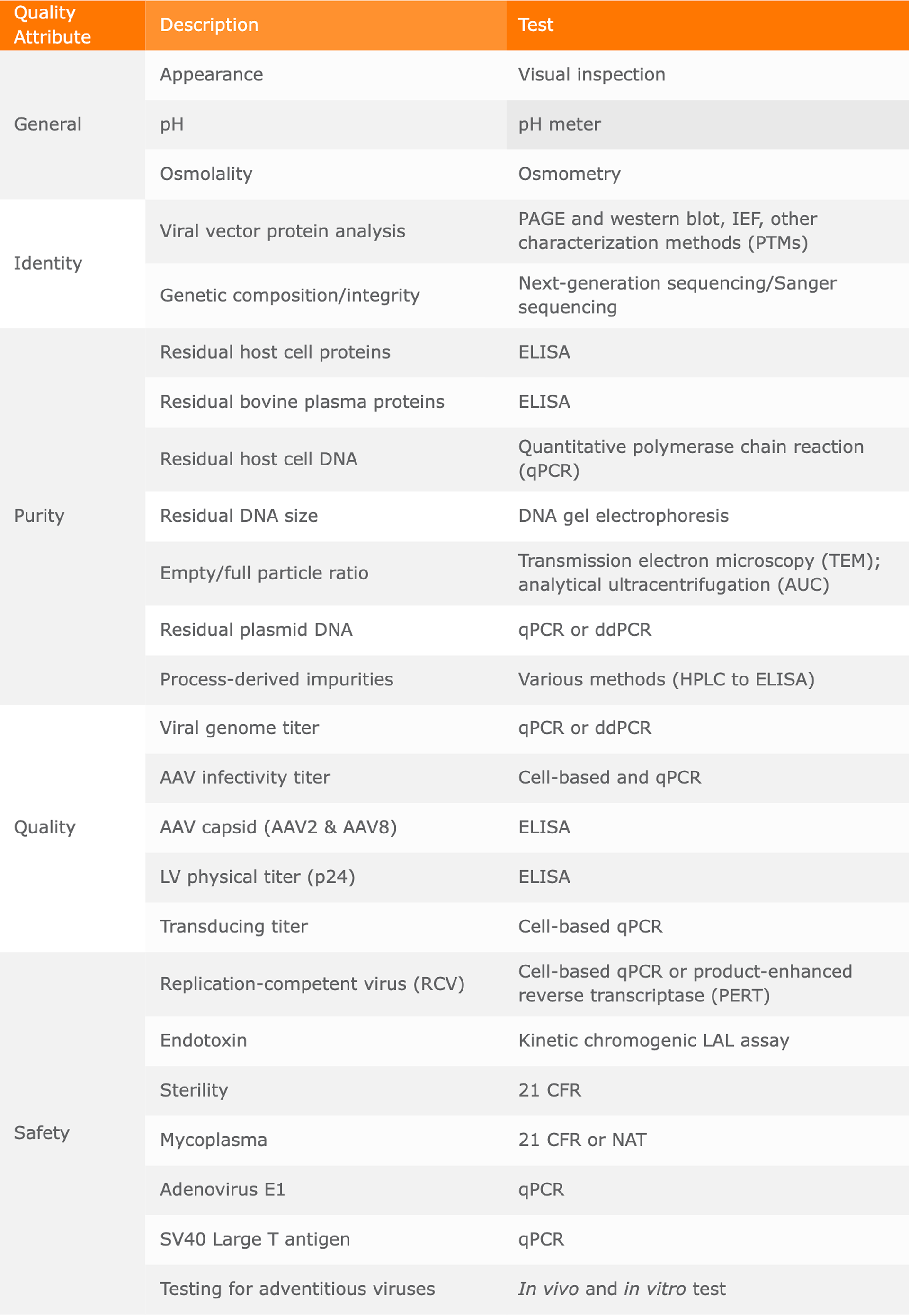
Table 11. Quality Control Assays for AAV Vectors
Source: Biovian[11]. Nice Insight, August 2024.
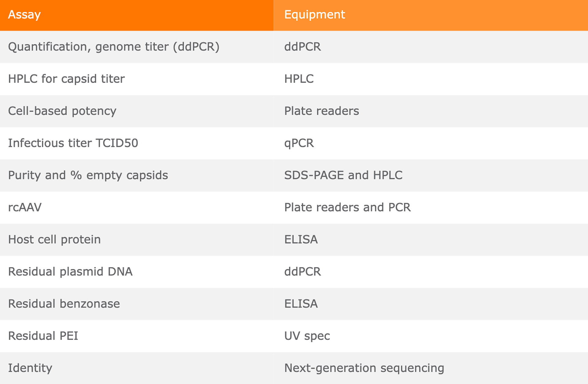
Table 12. Basic Analytical Equipment
Nice Insight, August 2024.
2. Analytics for LV Vectors
2.1. As with any biologic drug substance and drug product, raw material analysis, in-process monitoring, and product-release testing are essential to ensuring the quality and safety of LV vectors used for the production of genetically modified cell therapies.
2.2. The quality and purity of the plasmid DNA, culture media, transfection agents, and other raw materials must be confirmed. All starting materials and components must be qualified and suitable for the intended purpose. Analytics are required for in-process monitoring during cell culture, harvest, and each downstream unit operation. Product-release testing is necessary to confirm that LV vectors meet specifications required for use in the genetic modification of cells related to specific vector properties, impurity levels (e.g., HCPs, host cell DNA), and contaminants, such as mycoplasma and adventitious agents. CQAs for viral vectors include viral potency, identity, quantity, process residuals, aggregation, empty capsids, protein content, and product safety.
2.3. The complexity of LV and other vectors necessitates the use of numerous orthogonal analytical methods for complete characterization and quality determination.[12] Methods used for AAV vector analyses as detailed in Section 7.1 above are in general also applied to LV and other viral vectors.
2.4. Here again, the capsid viral proteins are crucial to the ability of LV vectors to genetically modify target cells; thus, full characterization of the capsid proteins and their purity is essential. To achieve this goal, the purity and structures of the plasmids used to produce the capsids must be confirmed. Similarly, the size, peptide sequence, and PTMs of the capsid, as well as its purity with respect to HCPs, host cell DNA and RNA, and partial and empty capsids, must be evaluated.
2.5. LV capsid protein analysis can be performed using traditional SDS-PAGE, immunoblotting, and ELISA techniques, as well as newer approaches, such as CE-LIF and LC-MS/MS. The capsid genome can be evaluated using PCR or high-throughput NGS, as well as newer, automated Sanger genome sequencing systems.
2.6. Viral vector impurities can also be rapidly identified and quantified using advanced LC-MS/MS and automated CE-SDS, in addition to or as a replacement for SDS-PAGE, ELISA, qPCR, and TEM. For determination of the full, partial, and empty capsids, techniques include NGS, spectrophotometry, electron microscopy, IEX chromatography, AUC, CE-LIF, and cIEF.
2.7. For rapid in-process monitoring of LV vector particles, HPLC has been recently reported to be effective.[13] The authors of that study developed a high-throughput method based on ion-exchange high-performance liquid chromatography (IEX-HPLC) for robust physicochemical characterization of LV particles in all types of samples, from crude LV supernatants to final purified products. This method was shown to be faster and more cost-effective than ddPCR and ELISA methods, due to its simple sample preparation and ability to track both total and functional vector particles. LV vector products must also be assessed for potency, typically using cell-based assays; physical titer, using methods such as the A260/280 assay and HPLC; and infectious or functional titer (expressed as PFU/mL or IFU/mL for AAV vectors), determined most often using the viral plaque assay, but also the TCID50 assay and the IFA.[14]
2.8. Potency is again difficult to assay, because different methods provide different results due to the need to measure biological activity in any number of specific cell types depending on the indication. But it is a crucial assay because it determines dosing. Assays for dose determination are product-dependent and must be optimized for the specific LV vector and qualified before initiating clinical studies, including demonstration of assay linearity, accuracy, precision, range, sensitivity, and specificity.
2.9. Safety testing is required for all viral vectors, just as it is for all biologics, including sterility, endotoxin, mycoplasma, adventitious virus, and replication-competent virus testing.[14]
2.10. Stability studies must be performed according to local and international regulations, including those established in the ICH guidelines and guidance published by the FDA[15] and the EMA.[16] They are necessary to ensure that the quality and activity of the LV vectors is not adversely affected during storage, such as due to the formation of protein aggregates. SEC and techniques such as field-flow fractionation multi-angle light scattering (FFF-MALS) and AUC are used for aggregate quantification.
3. Analytical Challenges
3.1. One of the remaining significant challenges to the development of gene and gene-modified cell therapies relates to the lack of analytics designed specifically for viral vector characterization. For AAV vectors, in addition to identity, viral titer, and other expected measurements, the ratio of full to partial and empty capsids must be determined, as the latter can lead to immunogenic responses. Detection of any capsids containing undesirable host cell or plasmid DNA is equally important, as they could potentially be toxic.
3.2. The goal for newer analytical techniques is to be able to apply them in a platform manner as simply as possible (such as no need for external standards) with little special skill or training required yet to still be able to obtain robust and reliable results independent of the operator and the laboratory.
3.3. Digital droplet PCR (ddPCR) is an attractive technique that has become widely used, eclipsing other quantitative PCR methods.[17] It provides absolute quantitation with higher accuracy sensitivity and precision but without the need for numerous amplification cycles or an external standard. It is being employed for the determination of genome integrity, vector genome titers, and infectious titer assays and quantification of residual plasmid, host cell, and unpackaged viral DNA, among other applications. Most vendors of ddPCR instruments offer kits designed specifically for cell and gene therapy applications.[18]
3.4. Cryogenic electron microscopy, meanwhile, is replacing more traditional transmission electron microscopy for characterizing viral vectors due to rapid advances in both hardware and software.[18] However, it still suffers from high costs, the need for skilled operators, and a relatively slow turnaround time (approximately one week).
3.5. Another form of advancement is the combination of multiple existing technologies into single instruments for more comprehensive and rapid analyses.[18] The Stunner instrument from Unchained Labs combines dynamic light scattering (DLS), static light scattering (SLS), and UV/Vis spectroscopy and can be used for a number of different assays, including determination of full/empty capsid ratios. The Aura device from Halo Labs uses fluorescence membrane microscopy (FMM) in combination with brightfield backgrounded membrane imaging (BMI) for rapid aggregation assessment of viral vectors and detection of subvisible particles in cell therapy cell cultures. Four label-free methods [DLS, SLS, back-reflection, and nano differential scanning fluorimetry (nanoDSF)] are combined in the Prometheus Panta device from NanoTemper for evaluation of thermal stability, genetic fill, and purity of viral vectors in a single analysis.[18]
3.6. New techniques are also being applied to viral vector analysis. In 2022, Refeyn introduced SamuxMP, a device using interferometric scattering microscopy (iSCAT), also referred to as mass photometry, to rapidly assess AAV full/empty capsid ratios for low-volume samples.[18] Charge-detection mass spectrometry is a single-particle variant of mass spectrometry that applies to heavy individual ions within heterogeneous samples.
3.7. Manufacture of viral vectors includes production of plasmids encoding helper-virus functions and the therapeutic gene; cell lines used to manufacture the vector, and other materials, followed by transfection; or the generation of mammalian or insect producer cell lines followed by infection, harvesting, purification, characterization, formulation, and fill/finish.
3.8. Viral vectors are much larger than recombinant proteins and antibodies and have been known to kill the cells used to produce them. Transient expression of the components essential to produce a virus also limits titers.
FAQs
What are the primary Critical Quality Attributes (CQAs) for viral vectors?
Critical Quality Attributes for viral vectors include viral potency, identity, quantity, process residuals, aggregation, empty capsids, protein content, and product safety. These attributes ensure the vector's ability to effectively deliver genetic material while minimizing immunogenic responses from impurities like host cell proteins (HCPs) or host cell DNA.
Why is the empty-to-full capsid ratio critical in gene therapy?
The empty-to-full capsid ratio is a vital purity metric because empty or partial capsids can trigger undesirable immunogenic responses in patients without providing therapeutic benefits. Conversely, capsids containing host cell or plasmid DNA are potentially toxic, making precise quantification via AUC, TEM, or cIEF essential for patient safety.
How does Digital Droplet PCR (ddPCR) improve viral vector manufacturing?
Digital Droplet PCR (ddPCR) improves manufacturing by providing absolute quantitation of vector genomes and residual DNA with higher accuracy and sensitivity than traditional qPCR. It eliminates the need for external standards or numerous amplification cycles, streamlining the determination of physical titers and genome integrity.
What analytical methods are used for AAV capsid protein characterization?
AAV capsid proteins are characterized using Capillary Electrophoresis (CE-SDS), Liquid Chromatography-Tandem Mass Spectrometry (LC-MS/MS), and Peptide Mapping via Q-TOF MS. These methods evaluate the peptide sequence, posttranslational modifications (PTMs), and purity, which are crucial for receptor binding, intracellular trafficking, and overall treatment efficacy.
What are the key differences between AAV and LV analytical requirements?
While both require safety, purity, and potency testing, Lentiviral (LV) vectors specifically require stability studies to prevent protein aggregation during storage. LV analysis often utilizes IEX-HPLC for rapid in-process monitoring and FFF-MALS or SEC for aggregate quantification, addressing the larger size and complexity of these vectors.
McCarthy, Sean, Zoe Zhang, and Fan Zhang. “Reproducible Peptide Mapping of Biotherapeutics with SWATH® Acquisition on the TripleTOF® 6600 System and BioPharmaViewTM Software.” Application note. SCIEX. 2017.

 Source: Biovian[11]. Nice Insight, August 2024.
 Nice Insight, August 2024.