Ultra-high definition is achieved through digital programming of colors using advanced nanotechnology.
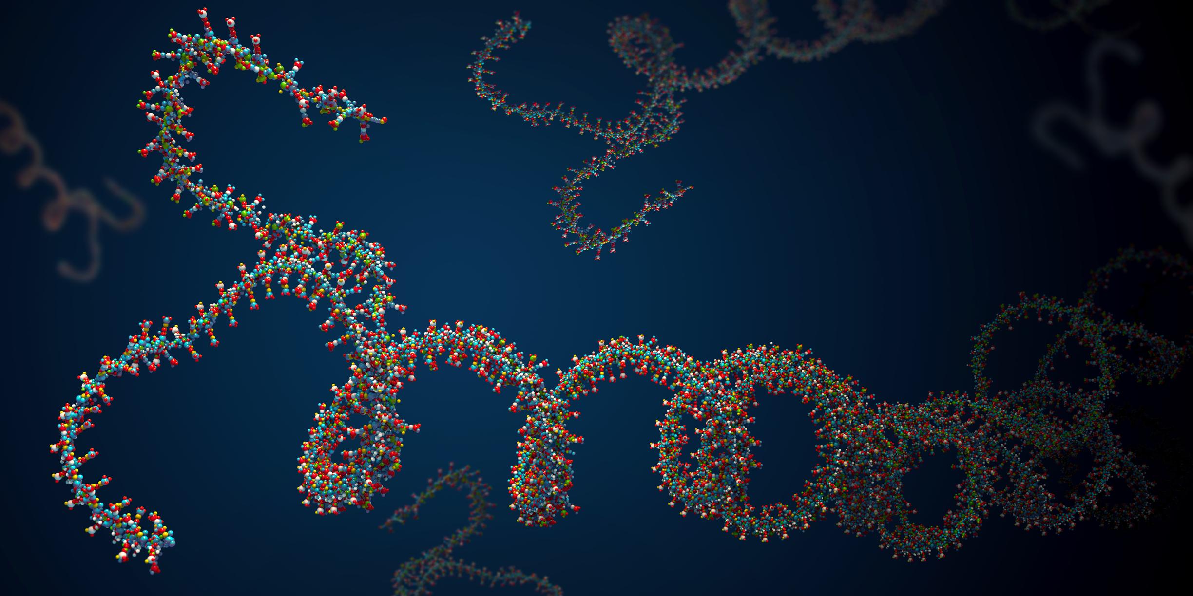
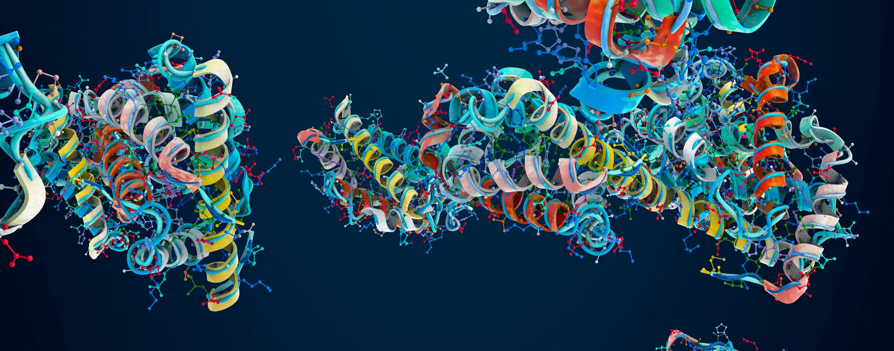
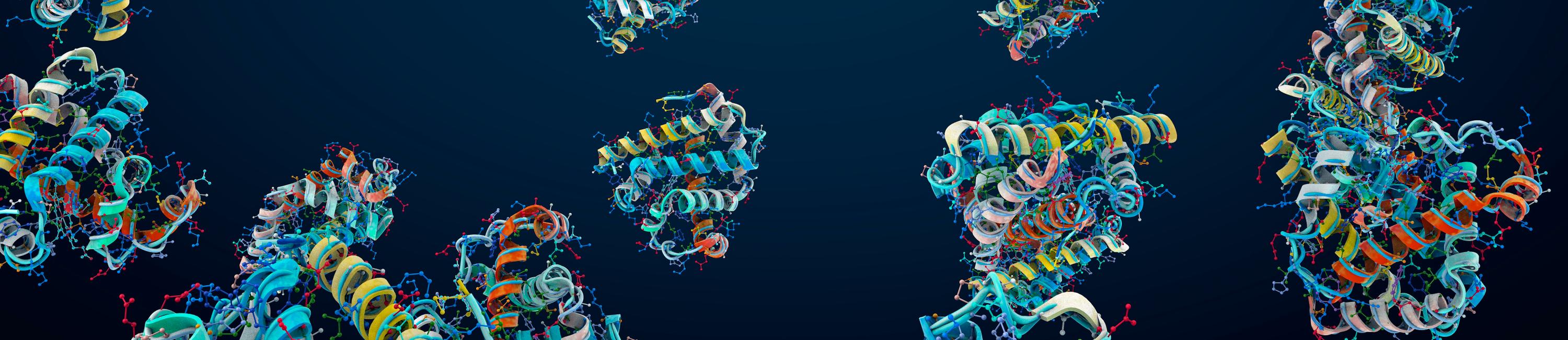
Scientists are able to obtain images of complex molecules, but have difficulty achieving the resolution necessary for simultaneously visualizing a large number of molecules as they mediate complex molecular processes. However, researchers at Harvard’s Wyss Institute for Biologically Inspired Engineering, the LMU Munich, and the Max Planck Institute of Biochemistry in Germany may have developed a solution. This team of scientists has integrated commonly used small fluorescent probes into self-folding DNA structures—such that their colors and brightness can be digitally programmed—yielding engineered and versatile metafluorophores. A total of 124 virtual colors can be used for microscopic imaging or other analytical methods, and hopefully going forward, will be modified to allow visualization of multiple molecules interacting with one another.
With conventional fluorescence microscopy, only a small number of molecules in a biological or clinical sample can be visualized simultaneously. The use of the metafluorophores, which are fluorescent dyes whose colors are determined by how their individual components are arranged in 3-dimensional structures, overcomes this limitation.
The DNA nanostructures are used as molecular pegboards, according to Peng Yin, a Core Faculty member at the Wyss Institute and Professor of Systems Biology at Harvard Medical School. Specific component strands are functionalized with one of three different fluorescent dyes at certain positions within the DNA nanostructure. With this approach, the team can generate up to 124 fluorescent signals with unique color compositions and intensities.
“Our study provides a framework that allows researchers to construct a large collection of metafluorophores with digitally programmable optical properties that they can use to visualize multiple targets in the samples they are interested in,” said Peng. The DNA nanostructures can then be used like a “barcoding system” for multiplexing to visually profile the presence of many specific DNA or RNA sequences in samples.
One challenge to using the larger DNA nanostructures was their limited mobility in thick tissue samples. To overcome this issue and also reduce the likelihood of non-targeted attachment of the DNA nanostructures, which would result in false fluorescent signals, the metafluorophores were designed to dynamically self-assemble from small component strands only after binding to the target, according to Ralf Jungmann, Ph.D., who co-conducted the study together with Yin and is a member of the faculty at LMU Munich and the Max Planck Institute of Biochemistry.
“These in-situ assembled metafluorophores can not only be introduced into complex samples with similar combinatorial possibilities as the prefabricated ones to visualize DNA, but they could also be leveraged to label antibodies as widely used detection reagents for proteins and other biomolecules,” Jungmann said.
Wyss Founding Director Donald Ingber, M.D., Ph.D., who is also the Judah Folkman Professor of Vascular Biology at Harvard Medical School and the Vascular Biology Program at Boston Children’s Hospital, as well as Professor of Bioengineering at the Harvard John A. Paulson School of Engineering and Applied Sciences, commented on the technology. “This new type of programmable, microscopy-enhancing DNA nanotechnology reveals how work in the Wyss Institute’s Molecular Robotics Initiative can invent new ways to solve long-standing problems in biology and medicine. These metafluorophores that can be programmed to self-assemble when they bind their target, and that have defined fluorescent barcode readouts, represent a new form of nanoscale devices that could help to reveal complex, multi-component, biological interactions that we know exist but have no way of studying today,” he said.