Accelerated ADC Development: The clinical and commercial success of first-generation Antibody-Drug Conjugates (ADCs) has triggered a massive pipeline of next-generation therapies, evolving from Paul Ehrlich’s "magic bullet" concept into a cornerstone of precision oncology.
The Three-Pillar Design Framework: Successful ADC engineering requires a synergistic balance between three critical components: the monoclonal antibody (mAb), the high-potency cytotoxic payload (HPAPI), and the chemical linker—including the specific conjugation chemistry used to bind them.
Precision Targeting & Systemic Stability: A primary goal of ADC design is maintaining stability in systemic circulation to prevent premature payload release, ensuring the toxic cargo is only "unlocked" once the antibody binds to specific tumor-associated antigens (TAAs).
Intracellular Payload Release: The efficacy of an ADC depends on its ability to be internalized by the target cell, where the linker must effectively detach the payload (often via lysosomal degradation or pH-sensitive cleavage) to trigger cell death.
Optimization of DAR: Modern drug discovery focuses heavily on the Drug-Antibody Ratio (DAR) and site-specific conjugation to reduce off-target toxicity and improve the therapeutic window for patients.
Originally Published: September 2024
1 A Short History Lesson
1.1 The success of therapeutic monoclonal antibodies has spurred interest in next-generation versions of the modality. One such innovation in the antibody space is the conjugation of a cytotoxic compound, the payload, to the antibody. The concept of a magic bullet — Zauberkugel — was first proposed by Nobel laureate Paul Ehrlich in 1907. The dream of developing a drug that will only injure the desired cells without harming healthy cells around it is finally becoming a reality. This reality takes the form of conjugating a toxic compound, the payload, to a monoclonal antibody. The road from Dr. Ehrlich’s 1907 proposal to today is a classic story of scientific progress in rapid leaps — and disappointing setbacks.
1.2 Researchers began in vitro studies with antibody–drug conjugates (ADCs) as early as the 1960s using polyclonal antibodies and radioisotopes and other cytotoxic drugs. The low drug–antibody ratio (DAR), combined with unstable linkers, led to poor pharmacokinetics and ultimately no therapeutics reached human testing. With the ability to create monoclonal antibodies, researchers could more precisely target the desired cells, which yielded more promising results.1
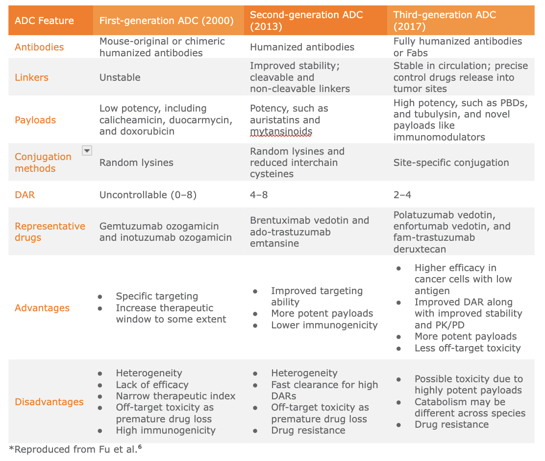
1.3 Human testing of ADCs began in 1983 with the investigation of a monoclonal anti-carcinoembryonic antigen antibody conjugated to vindesine.2 Vindesine is a synthetic alkaloid that induces mitotic arrest.3 This drug was not successful, however, and it wasn’t until 2000 before an ADC was approved by the FDA. This drug, Mylotarg, was developed by Pfizer to treat blood cancers and represents the paradigm of first-generation ADCs because the payload, ozogamycin, is bound covalently to lysine residues in the antibody and cannot be cleaved.4 Mylotarg was voluntarily withdrawn from the market in 2010 due to high off-target toxicity and was reintroduced to the market in 2017.5,6 A summary of the evolution of ADC development is presented in Table 1.
1.4 The first generation of ADCs linked a traditional chemotherapy agent such as methotrexate, vinblastine, or doxorubicin via a non-cleavable linker. They were conjugated through either lysines or cysteines on the molecule, which yields a higher variability in the DAR than what is desired. The low efficacy of the first-generation ADCs prompted a shift toward novel and highly potent active pharmaceutical ingredients (HPAPIs), such as the tubulin inhibitor maytansine, that can achieve up to 100 to 1,000 times greater potency. The drawback of using HPAPIs as a systemic therapy, however, is the increased incidence of severe adverse events such as neurotoxicity and gastrointestinal reactions. Ultimately, the side effects of systemically delivered HPAPIs prompted renewed interest in conjugating these potent compounds to an antibody to create highly targeted drugs.7
1.5 Kadcyla, developed by Genentech/Roche and approved by the FDA in 2013, is considered to be the first of the second-generation ADCs. It is a treatment for HER2-positive breast cancer and is the first ADC to reach $1B in sales. Kadcyla is also notable because it demonstrated the success of targeting solid tumors with an ADC. Adcetris (Seagen) and Zynlonta (ADC Therapeutics) are also classic representatives of the second-generation class of ADCs. Overall, second-generation ADCs are characterized by their high-quality monoclonal antibody, conjugated via lysine or cysteine to an HPAPI via a non-cleavable linker. This generation of drugs still suffers from variable DAR and low stability of the linker in blood, as well as a relatively high incidence of toxic side effects.7
1.6 Classic examples of third-generation ADCs include Besponsa (Pfizer, approved in 2017), Enhertu (Daiichi Sankyo, approved in 2019), and Padcev (Seagen, approved in 2019). These therapies enjoy better stability and pharmacokinetics, in part because they all use a cleavable linker.7
1.7 Interestingly, while the first ADC launched was for a hematological target, the number of ADCs with solid tumor targets have outnumbered the former. The pipeline of ADCs in clinical trials, as discussed in detail in the next section, shows a higher prevalence of ADCs targeting solid tumors as well. Looking at the category of payloads used in the approved ADCs, most use tubulin-binding agents, but we see an increase in the prevalence of topoisomerase-I inhibitors in the upcoming ADCs. With respect to the linkers used, there is a clear majority of cleavable linkers over non-cleavable linkers. The upcoming generation of ADCs has improved characteristics with breakthroughs in payloads, antibodies, linkers, and the base technologies.
Table 1. Evolution of ADCs*
2 Intro to an Antibody–Drug Conjugate
2.1 The conjugation of an antibody to a toxic payload, resulting in an antibody–drug conjugate (ADC) has potential to deliver cytotoxic compounds with the precise specificity of an antibody. Because delivery can be precise at the cellular level, these therapeutics have the potential to greatly reduce the dose required of the cytotoxic compound while also reducing the effects of off-target toxicity.
2.2 Although the name implies an antibody is the key biologic portion of such compounds, any biologic with potential to bind precisely to its target within the host can serve as the conjugated partner. For convenience, we will use the term “ADC” throughout this report, although most of the concepts also apply to other biologics, such as soluble receptors and RNA. Likewise, the cytotoxic partner in the ADC can be any type of payload. Small molecules are the most prevalent payload class, but immunotoxins, siRNA, and cytotoxic peptides are also becoming more common in clinical trials. A simple illustration of a classic ADC is presented in Figure 1.
2.3 If we are considering the payload to be exclusively a cytotoxic compound, then it follows that the therapeutic indications are those in which death of certain cell types is the desired result. Indeed, most ADCs are indicated for cancers, but they are also in development for bacterial infections and atherosclerosis.8
2.4 Activity of the ADC depends on the cellular uptake of the drug via phagocytosis, where the payload is active. An ideal ADC would have the following characteristics:4
A specific targeting moiety (such as an antibody or equally specific biologic) that will bind tightly to the target but not other types of cells. The advanced state of cancer research and antibody discovery technologies aids the design of this part of the drug. Biosimilar antibodies can be employed and have the advantage of well-understood manufacturing and clinical characteristics.
A potent cytotoxic compound that is active inside a cell, but will not have adverse effects in the bloodstream. Any number of mechanisms to induce cell death can be employed. Some payloads are designed to be inactive in serum or otherwise masked until the drug is phagocytosed.
A linker that will bind effectively to both the biologic targeting moiety and the cytotoxic payload. The ideal linker will be stable in serum, but can be or may be designed to be cleaved inside a cell. Manufacturing considerations are of paramount importance in linker design.
2.5 Antibodies are covered in many other resources, including our own Mammalian Cell Culture, Biologics: 2024 Market Insight, CDMO Pricing and Competitor Benchmarking report, so we will not devote additional space to this component of ADCs here.
Figure 1. Structure of an ADC
The basic structure and nomenclature of an antibody–drug conjugate are presented. The linker can be fixed or cleavable, as discussed in the text. The payload can be any cytotoxic entity, such as a small molecule, small peptide, oligonucleotide, protein toxin, or radioisotope. Payloads can be conjugated to the antibody anywhere on the molecule, but conjugation in the ligand-binding region (represented in yellow) is undesirable.
3 Payloads
3.1 Antibody-based therapeutics have enjoyed incredible success in part due to their precise and effective binding capabilities. They can serve as effective flags to highlight tumors and present tumor-associated antigens (TAAs) to the patient’s immune system, but can be limited in their efficacy, however, especially in cases where the immune system is depressed by disease or even the tumor itself.
3.2 There are a number of cytotoxic compounds that have been used as systemic chemotherapies, but their side effects can be dramatic. Linking the chemotherapy agent to a precise targeting moiety such as an antibody, therefore, has potential to take advantage of the best features of both classes and reduce the side effects of both.
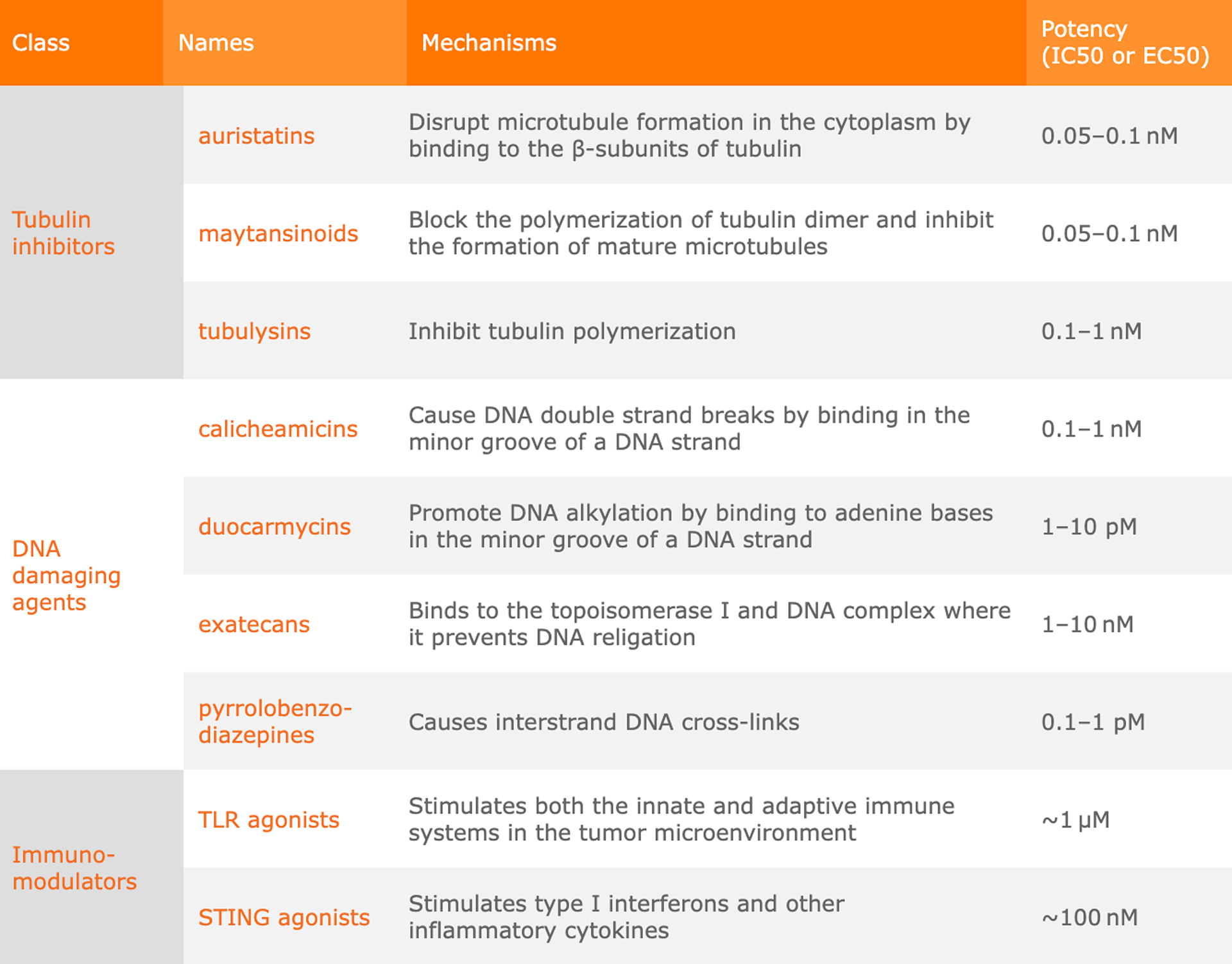
3.3 The ideal payload characteristics are presented in Table 2. The cytotoxic payload of an ADC can be a small molecule, immunotoxin, radioisotope, or any other cytotoxic compound. Maiti et al.9 provides a useful review of some common payload activities.
Table 2. Features of an Ideal ADC Payload7
Table 3. Common ADC Payload Categories7
4 Conjugation Technologies
4.1 Conjugation technology is, of course, intimately tied to payload and linkers in the ADC. We have summarized some advantages and disadvantages of different technologies in Table 3, and have provided some schematics of different conjugation methods in Figure 2 and Figure 3.
4.2 In the most simple methods of conjugation, linkers are often conjugated to available lysines or cysteine residues on the protein. Lysines are often conjugated using amide coupling to an active carboxylic acid ester on the linker while cysteine residues are conjugated to the linker via disulfide bridges. The interchain cysteine residues present in most antibodies serve as convenient conjugation points because they are usually surface-exposed.
4.3 One disadvantage to each of these stochastic methods is the potential for heterogeneity in the number of payloads per antibody. There are typically about 40 available lysines and two to eight cysteines that can be made available, although the number of each type of residue can be modified by changing the peptide sequence. Even if a small number of target residues are available, the variability in number of payloads conjugated to the antibody is still higher than desired. Conjugation via cysteine residues is currently the most common method employed in the ADC pipeline because it has a reasonable balance between low drug–antibody ratio (DAR) variance, convenience, and serum stability and is often part of site-specific conjugation methods.10
4.4 If the desired conjugation residue is in the antigen-binding domain, the payload may interfere with antibody targeting. This hurdle is readily overcome with alternations to the peptide sequence, creating a simple site-specific conjugation method.
4.5 One notable site-specific conjugation technology based on cysteine conjugation is THIOMABTM, developed by Genentech. The technology conjugates a polymer holding a large number of payloads via cystine reduction. Using this technology, high and consistent DARs have been achieved, yet the platform is flexible enough to allow conjugation of many types of payloads to nearly any antibody.4,11,12
4.6 When cysteine residues are either not available in the sequence or not desired, researchers must look for alternate conjugation sites. Recent advances in technology to incorporate nonnatural amino acids into recombinant products open up an opportunity to introduce new sites and new chemistries. In these cases, however, the conjugation may be more straightforward than incorporation of the nonnatural amino acid. Researchers also have to pay special attention to the immunogenicity and hydrophobicity of the nonnatural residues within the molecule.4,13
4.7 Enzymatic ligation can enable highly specific conjugation chemistry if the appropriate peptide sequence is incorporated into the antibody. Other methods of site-specific conjugation that the reader may want to explore further are glycoconjugation14 and pClick technology.15
Table 4. Common ADC Conjugation Technologies4
Figure 2. Schematic Diagrams of Stochastic Conjugation Technologies
A simple representation of the methods for lysine conjugation (top panel) and cysteine conjugation (bottom panel) is shown. Although only one payload is represented (blue dot), ADCs conjugated in this manner typically carry several payloads per antibody. Reproduced from Fu et al.4
Figure 3. Schematic Diagrams of Common Site-Specific Conjugation Technologies
A simple representation of the methods for some common site-specific conjugation technologies is shown. As described in the text, many of these technologies ensure that only two to four payloads are conjugated to each antibody. Reproduced from Fu et al.4
5 Linker Technologies
5.1 The biologic portion of an ADC must be chemically bound to its payload through a linker. These short peptides or small molecules can be attached to the antibody and payload in a variety of chemistries with a range of different effects. It helps to classify them as cleavable or non-cleavable (Table 5 and Figure 4).
Non-cleavable linkers
5.2 The first linkers used in ADC development are small molecules which are conjugated via covalent bonds to the antibody and the payload. Typically, there are about 10 free lysines on an antibody and two to eight cysteines that could either be made available chemically or engineered into the protein sequence.16 Because the payload is covalently bound to the antibody, dispersion of the payload depends on the uptake of the ADC into the cellular cytoplasm or lysosomes, where low pH or endogenous proteases will cleave the bonds and free the payload from the antibody. Because release of the payload depends entirely on the physiology of the cell in which it is phagocytosed, ADCs with non-cleavable linkers have a higher rate of off-target effects.16
5.3 ADCs designed with non-cleavable linkers, however, have an advantage in that they are typically more stable in serum and have been demonstrated to have superior efficacy with some therapeutics.10
Cleavable linkers
5.4 As the name implies, cleavable linkers are designed to degrade and release their payload under specific circumstances, which generally fall into three categories: low pH, reducing environments, and specific enzymatic reactions. Because the cleavage is specifically designed, cleavable linkers should attain lower rates of off-target effects.
5.5 The three common classes of cleavable linkers are as follows:
Acid-cleavable linkers are cleaved by hydrolysis in the low pH of the tumor lysosomes, yet they are stable at pH 7 of serum. Chemical triggers that are dependent on acid cleavage include hydrazones, carbonates, and silyl ethers.17
Reducible linkers are cleaved in a glutathione-dependent manner in the tumor cytoplasm. Tumor cells express glutathione and other disulfide isomerases in higher concentrations than healthy cells so the likelihood of activity inside healthy cells is diminished.10
Enzymatic-cleavable linkers are only cleaved in the presence of their cognate enzyme. Enzymes that catalyze cleavage can be proteases (such as cathepsin), glycosidases (such as β-galactosidase and β-glucuronidase), or phosphatases. These enzymes are typically found only in lysosomes so the risk of release outside of a cell is greatly reduced.10,17
Table 5. Overview of ADC Linker Classes10
Figure 4. Types of Linkers used in ADCs
The types of cytotoxic payloads and their relationship to other payload categories are shown.
6 Imaging and Diagnostic Applications
6.1 Antibodies and ADCs have utility outside of therapeutics. Antibodies, often conjugated to some other entity, have long been used as critical research reagents for basic assays such as enzyme-linked immunosorbent assays (ELISAs), Western blots, flow blots, and flow cytometry.19 Antibodies and ADCs are also becoming increasingly critical in imaging applications, such as immunoPET and biopsies.
6.2 ImmunoPET uses radiolabeled antibodies to precisely visualize the cancer, which is especially critical when cancers have metastasized. Doctors can’t biopsy all of the possible metastatic sites, so they use specific imaging to understand the location and nature of the tumors. Imaging biologics are linked to short-lived radiolabels such as 89Zr or 124I.18 This type of imaging can support diagnosis as well as monitor therapeutic efficacy over time.
6.3 Precise molecular labeling is also useful in biopsy diagnosis, and the technique doesn’t always require radioisotopes. For example, tagging an antibody with fluorophores that change color when taken up by the cell can inform clinicians about the metabolism of a tumor in vitro as well as the efficacy of a therapeutic.19
6.4 Whether the clinician is using in vitro or in vivo imaging, a particularly useful aspect of this technology is the potential to label one biologic with different payloads that have different applications. If a researcher uses an antibody to image a tumor, not only can the physician determine if there is cancer and where it is located they can also more readily gauge the potential efficacy of the antibody when it is used as a therapeutic, either unlabeled or conjugated to a cytotoxic payload. For this reason, our clinical trial analysis includes both therapeutic and diagnostic antibody–drug conjugates.
6.5 As imaging technologies become more sophisticated, sensitive, and readily available, they are being implemented more often as part of clinical treatment plans of all types. For this reason, the molecular imaging market, already estimated to be near $5 billion in the U.S., is predicted to reach $15 billion by 2033.20
FAQs
What is the "Magic Bullet" concept in cancer therapy?
The "Magic Bullet" or Zauberkugel is a medicinal concept proposing drugs that selectively target and kill diseased cells without harming healthy tissue. First envisioned by Paul Ehrlich in 1907, this dream is realized through Antibody-Drug Conjugates (ADCs), which use monoclonal antibodies to deliver cytotoxic payloads directly to specific cellular targets.
How do first-generation and third-generation ADCs differ?
Third-generation ADCs offer superior stability and pharmacokinetics compared to first-generation versions by utilizing cleavable linkers and site-specific conjugation. While early ADCs like Mylotarg relied on non-cleavable bonds and random lysine/cysteine residues—leading to variable drug-antibody ratios (DAR)—modern iterations achieve precise control over payload release.
What are the main components of an Antibody-Drug Conjugate?
An ADC consists of three primary components: a monoclonal antibody (the targeting moiety), a potent cytotoxic payload, and a chemical linker. The antibody homes in on specific tumor-associated antigens (TAAs), while the linker ensures the payload remains stable in systemic circulation before releasing its toxic cargo inside the target cell.
Why is the Drug-Antibody Ratio (DAR) important for ADC efficacy?
The Drug-Antibody Ratio (DAR) determines the potency and safety profile of the therapeutic by defining the average number of payload molecules attached to each antibody. Low DAR can lead to poor efficacy, while excessively high or variable DAR often results in rapid clearance from the blood and increased off-target toxicity.
What is the role of cleavable linkers in reducing side effects?
Cleavable linkers reduce off-target toxicity by ensuring the cytotoxic payload is released only under specific intracellular conditions, such as low pH or enzymatic triggers. Unlike non-cleavable linkers, these are designed to remain stable in the neutral pH of the bloodstream but degrade rapidly within tumor lysosomes.
How are ADCs used beyond cancer treatment?
While primarily used in oncology, ADCs are increasingly utilized in diagnostic imaging and the treatment of bacterial infections or atherosclerosis. For example, immunoPET employs radiolabeled antibodies (using isotopes like 89Zr) to visualize metastatic sites, allowing clinicians to monitor therapeutic efficacy and metabolism in vivo
 2 Intro to an Antibody–Drug Conjugate
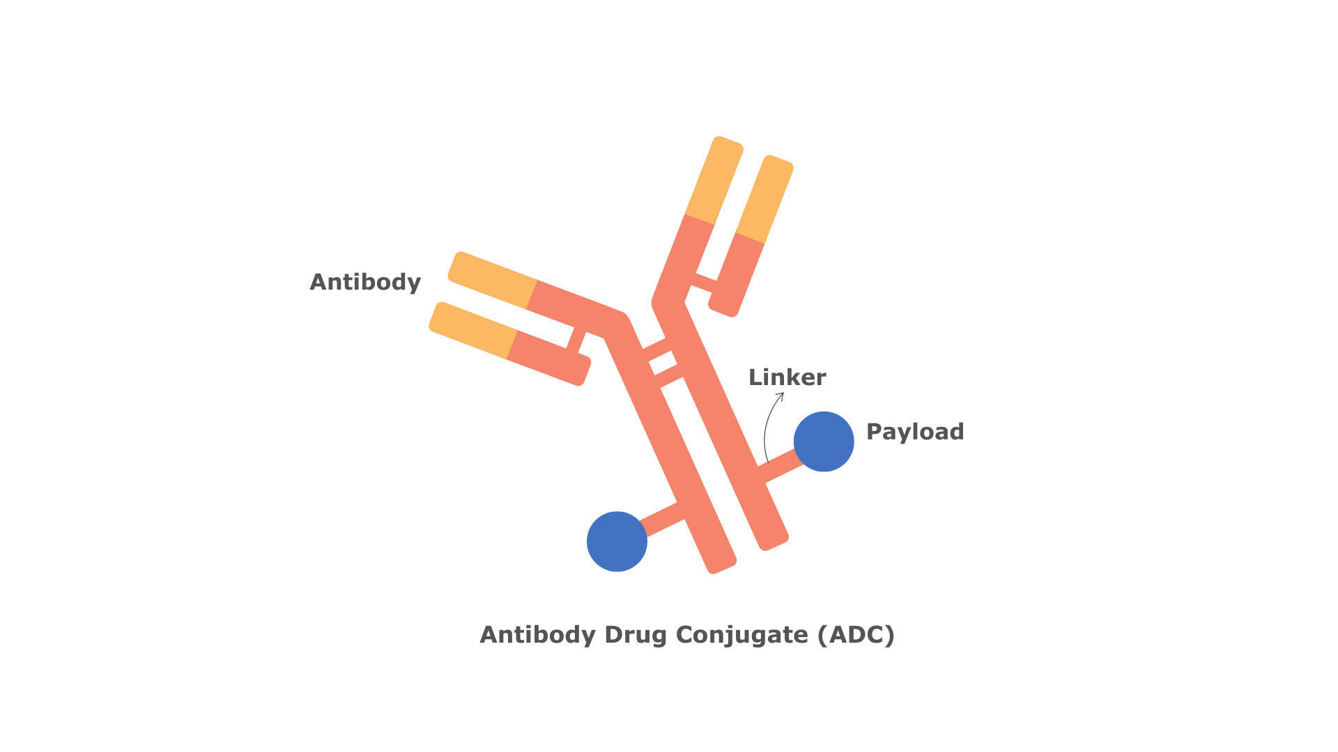 Figure 1. Structure of an ADC
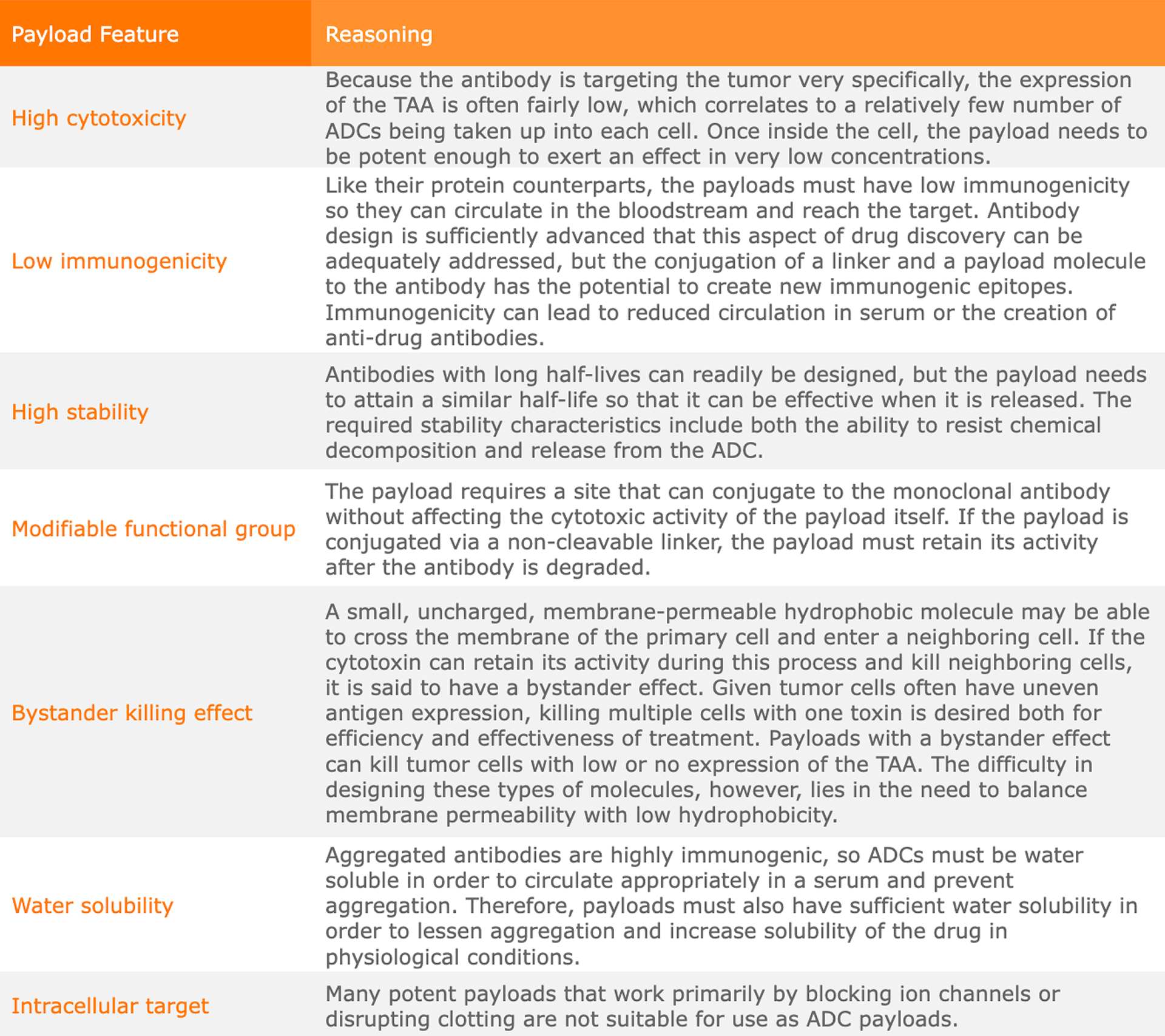 Table 3. Common ADC Payload Categories7
 4 Conjugation Technologies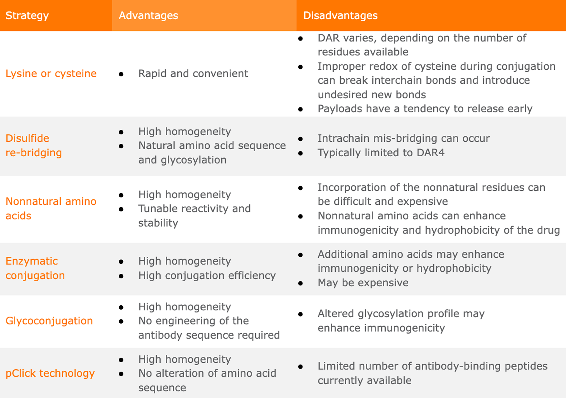
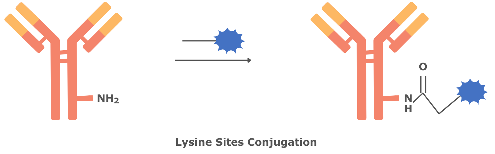
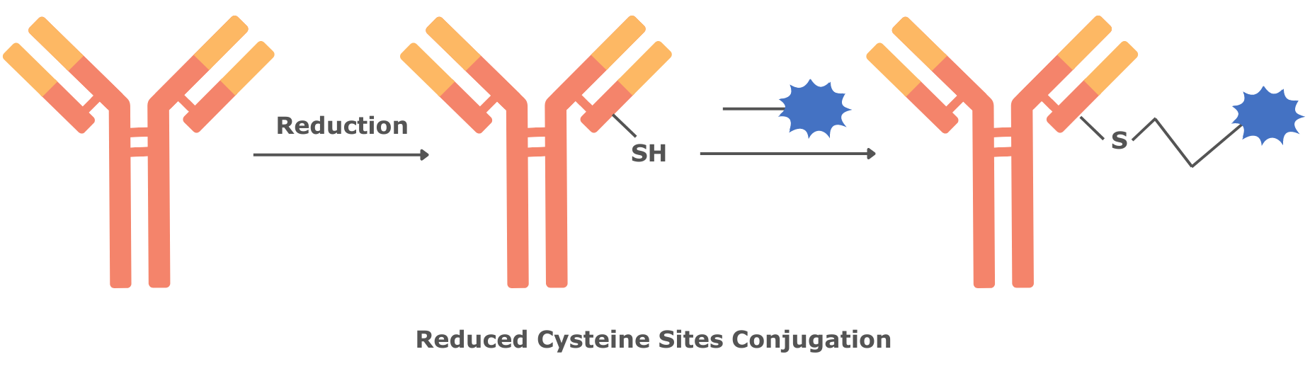 Figure 2. Schematic Diagrams of Stochastic Conjugation Technologies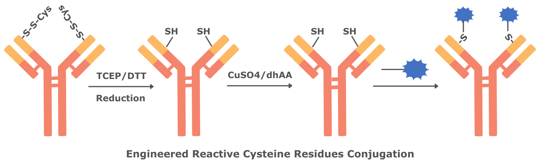
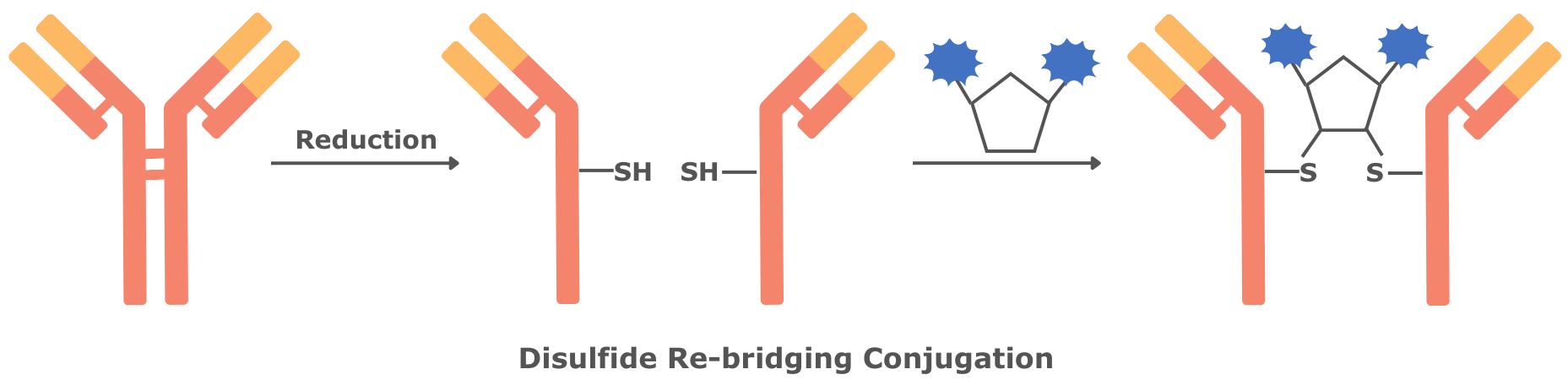
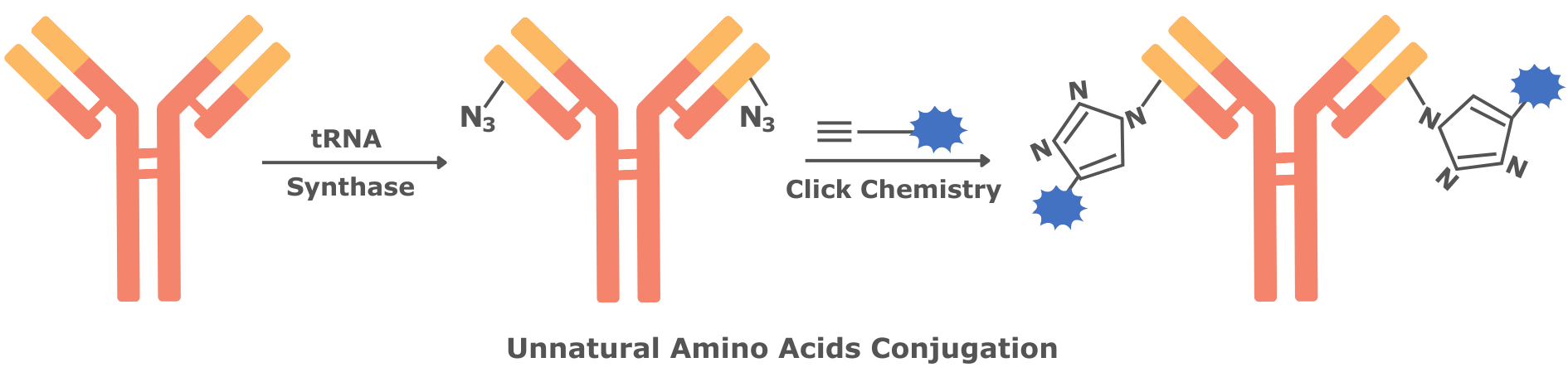
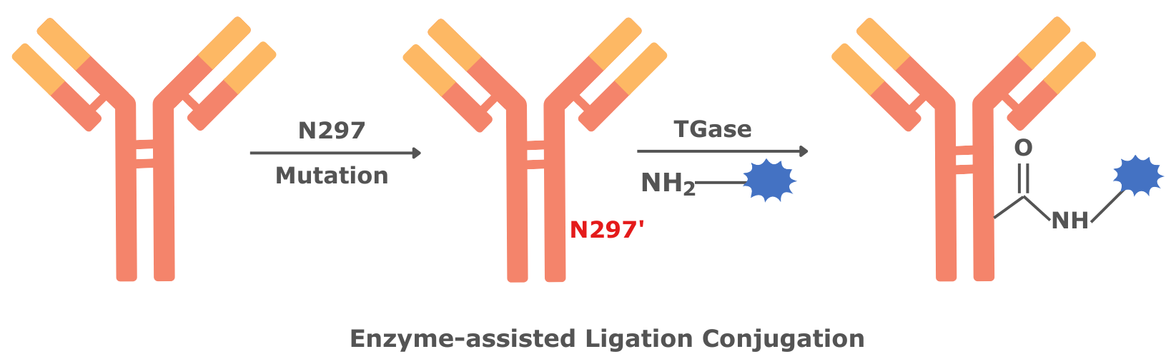
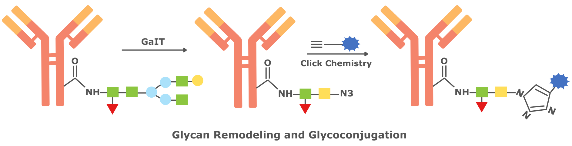
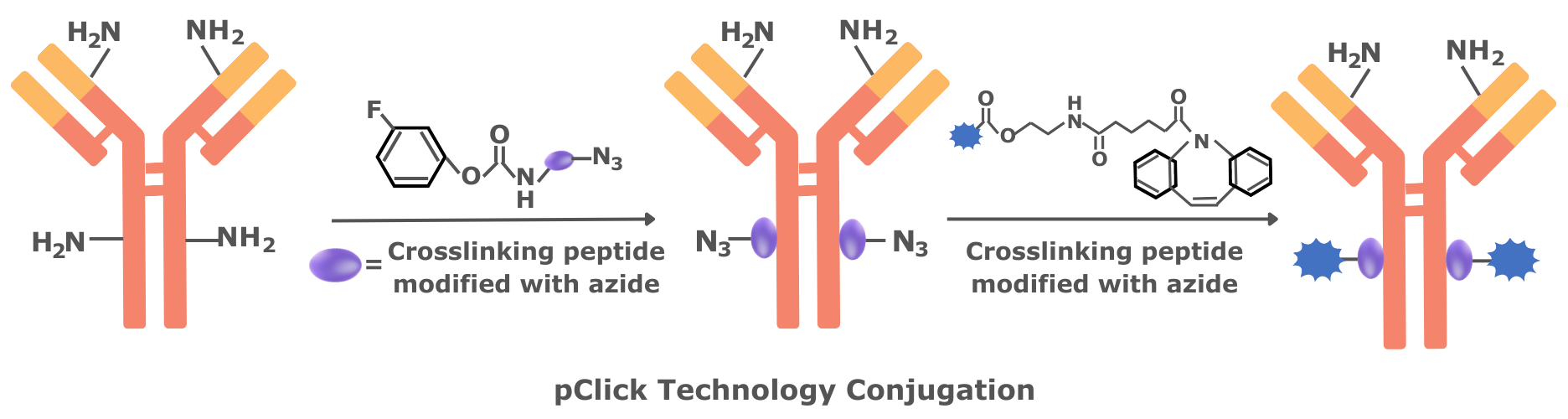 Figure 3. Schematic Diagrams of Common Site-Specific Conjugation Technologies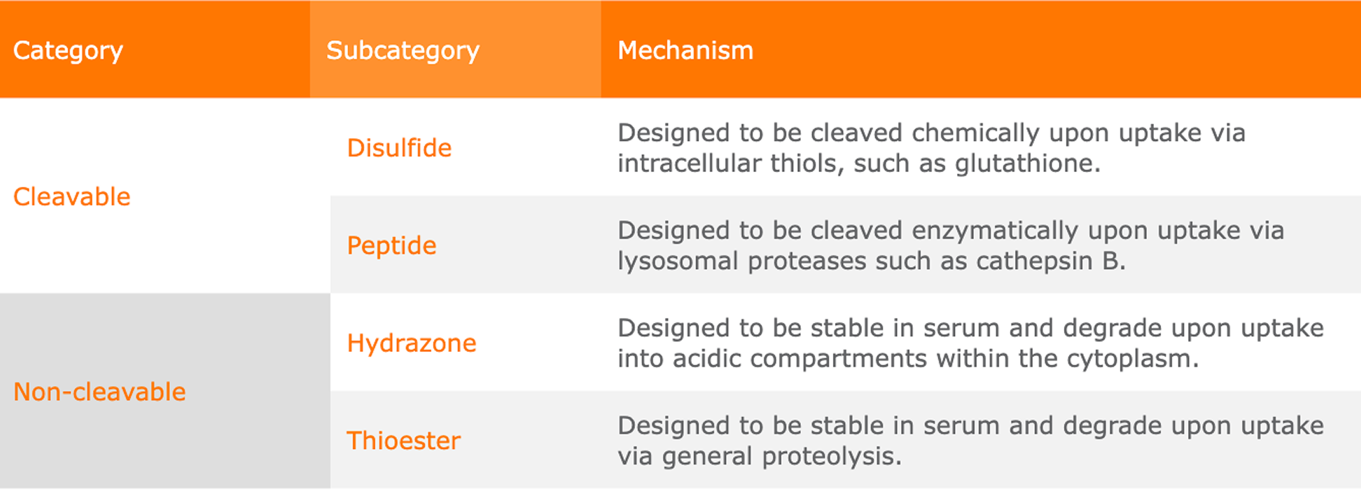
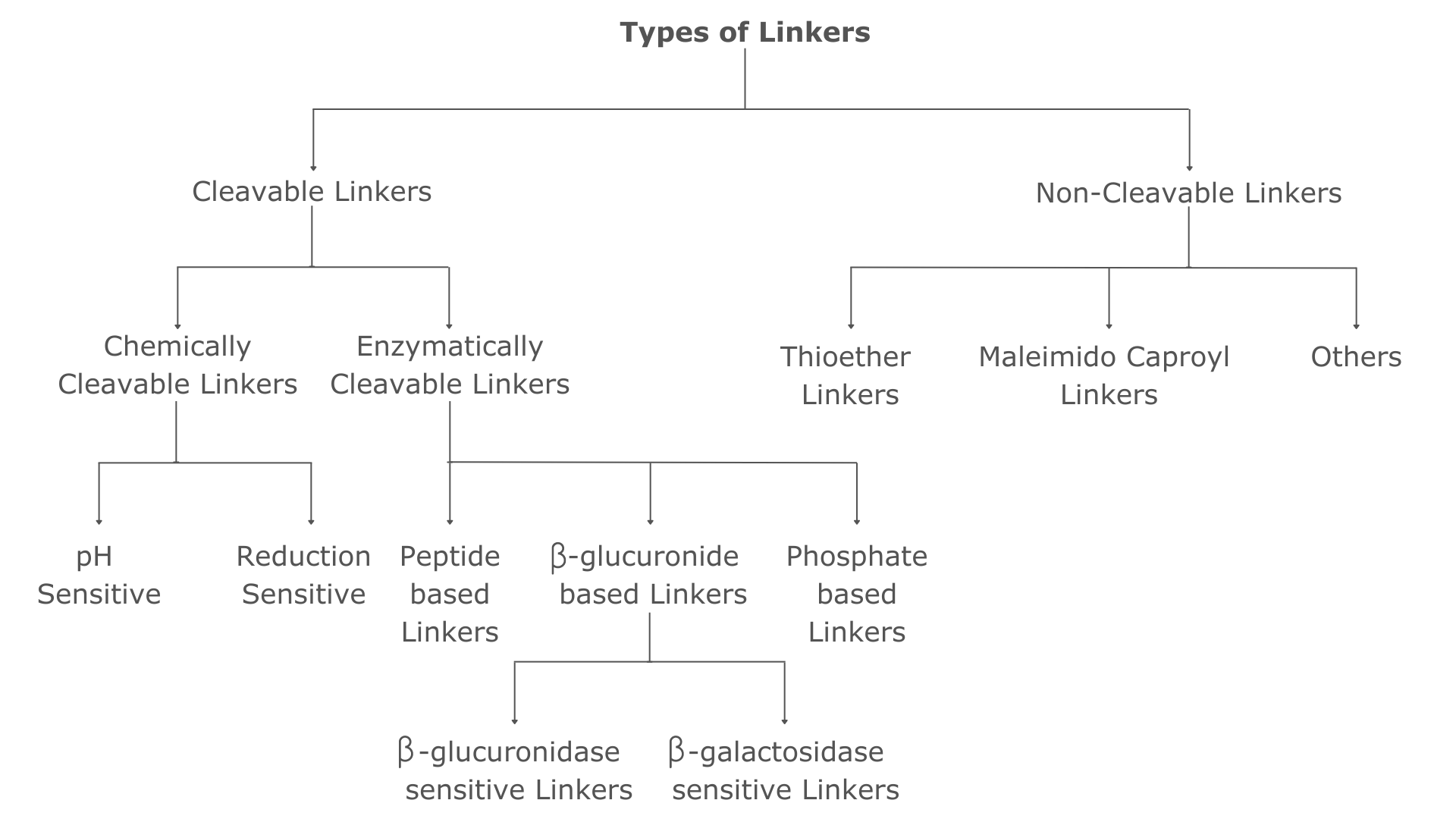 Figure 4. Types of Linkers used in ADCs