Protease Stability & Efficacy: The therapeutic success of a monoclonal antibody (mAb) is heavily influenced by its structural integrity—specifically how the molecule handles cleavage by standard proteases.
Engineering "Frankenbodies": By rearranging the natural domains of the traditional Y-shaped antibody, developers can create innovative formats like scFvs and bispecific antibodies to unlock new therapeutic mechanisms.
Design with Manufacture in Mind: Creating a "frankenmolecule" requires more than just biological creativity; designers must account for complex cell line development and manufacturing hurdles during the initial formatting stage.
Strategic Discovery Routes: Choosing a bispecific format isn't just a structural decision—it fundamentally dictates the antibody discovery path and the screening technologies required.
A Holistic Development Approach: For next-generation therapeutics to succeed, developers must evaluate discovery, development, and large-scale manufacturing alongside therapeutic properties before the design phase begins.
1 Anatomy of an Antibody
1.1 Most readers of our publishing platform will be familiar with the basic structure of an antibody, but we will review the concepts here for those who are new to the field.
1.2 Antibodies are Y-shaped heterotetramers secreted from B cells. The three sections of the Y are loosely connected by a flexible linker region that contains disulfide bonds (hinge region). The stem of the Y is a conserved region (constant region), while the tips of the Y vary between molecules (variable region) and bind to antigens (Figure 1).1
1.3 Each antibody consists of four polypeptide chains. The larger one, the heavy chain, is about 50 kilodalton (kDa), while the other, the light chain, is about 25 kDa. Two copies of each chain are bound together in a fully formed antibody. A typical antibody is about 150 kDa, and this basic structure is common to all five classes of natural antibodies (IgA, IgD, IgG, IgE, IgM).
1.4 While each antibody consists of identical copies of the heavy and light chain, there are different isotypes of each. Two classes of light chain, denoted κ and λ, have been identified, although no difference in function has been identified. The relative ratio of κ:λ in humans is 2:1, and variation on this ratio can indicate abnormal proliferation of a single B cell. An excess of identical light chain in a person might indicate a B cell tumor.
1.5 There are five isotypes of heavy chain denoted α (IgA), δ (IgD), γ (IgG), ε (IgE), and μ (IgM). There are four subclasses of γ, denoted IgG1, IgG2, IgG3, and IgG4. IgG are the most common class of antibodies found naturally, and are the most common basis of therapeutic antibodies. These distinctions of the IgG subclass are important for drug developers who are designing the specific effector function for their therapeutic.
1.6 The amino-terminal region of heavy and light chains, approximately 110 amino acids, is variable between antibodies, designated VH (variable region on the heavy chain) and VL (variable region on the light chain). The remaining carboxy-terminal region is constant, designated CH and CL. Multiple CH domains exist and are designated CH1, CH2, and CH3 (Figure 1).1
1.7 Antibodies can be readily cleaved by different proteases in predictable patterns. Two, in particular, are important for discussions of the novel antibody-based formats in the next section. Partial cleavage with papain separates the heavy chains such that three fragments are created. The CH2 and CH3 fragments of the two heavy chains remain bound together by their disulfide bond, and are designated the constant fragment (Fc). (Fc actually stands for crystallizable fragment.) The variable regions are then freed in two identical antigen-binding fragments (Fabs). Pepsin, however, cleaves the antibody such that CH2 is degraded and only a portion of the Fc (designated pFc') remains. This cleavage pattern also leaves the two Fab regions linked to each other, designated F(ab')2 (Figure 2).1
Figure 1. Structure of an Antibody
The basic structure and nomenclature of an antibody are given in the top panel. In the lower panel, the domain structure of the peptide chains in an antibody is given. Numbers designate approximate amino acid residues that correlate to the domains within each protein.
Figure 2. Common Antibody Fragments Created by Protease Cleavage
Common antibody fragments created by cleavage of papain and pepsin are shown.
2 New Antibody Formats
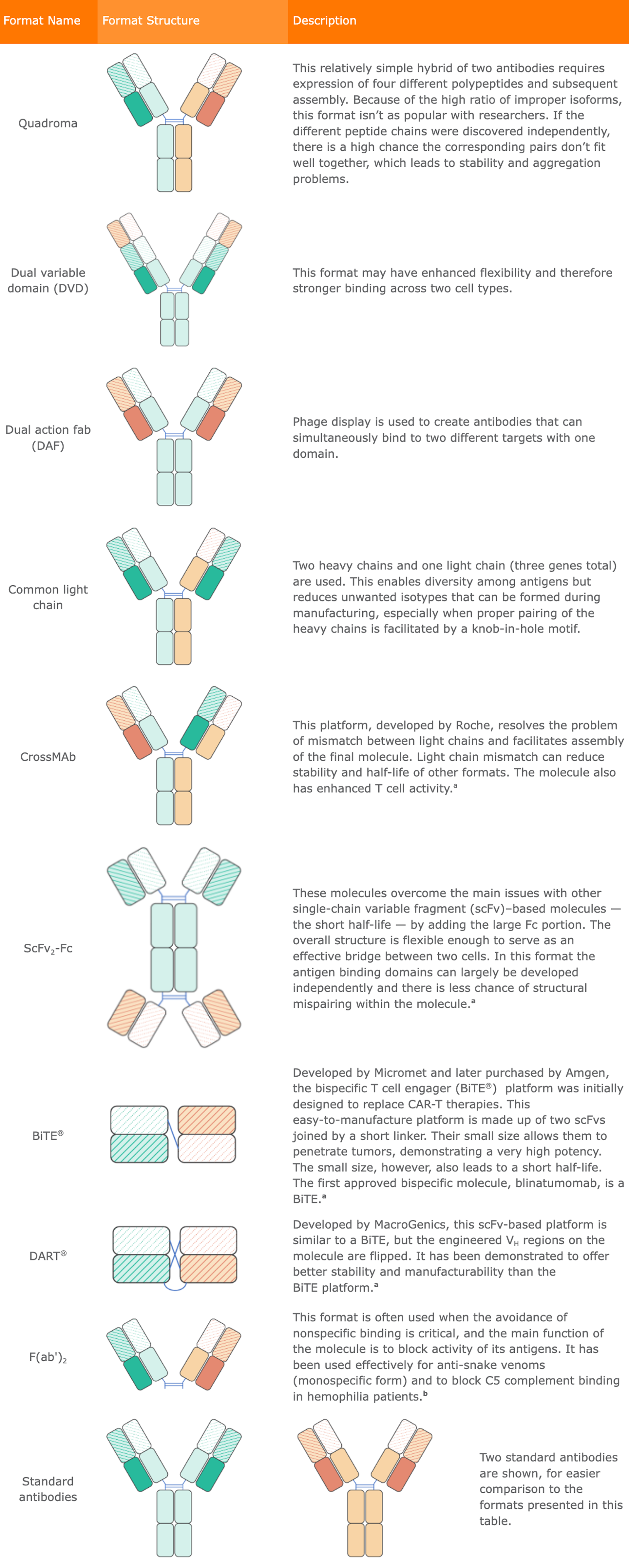
2.1 The widespread success of antibody therapeutics has encouraged development of variations on the form. Antibodies that could specifically bind to two different targets (bispecifics) were the first variation to gain traction in the market. The first bispecific antibody (bsAb) was approved by the FDA in 2014. Blinatumomab (brand name Blincyto, Amgen) is used to treat relapsed or refractory B cell precursor acute lymphoblastic leukemia (ALL). Its mechanism of action is to bind CD3 on T cells and CD19 on the tumor B cells. Since its approval, many more drugs that function by bringing the cytotoxic CD3+ T cell in contact with the tumor have been approved, and are classified as T cell engagers. The concept of bispecific T cell engagers (BiTEs) has since expanded to the development of natural killer (NK)–cell engagers and macrophage engagers. There are over 40 different bispecific formats in development. A few of the most common formats are presented in Table 1. In general, the smaller fragments can self-assemble and therefore can be easier to manufacture. The smaller structures may also enable conformational flexibility that enhances formation of the bridge between two cells. The smaller formats, however, have shorter half-lives. Blinatumomab, for example, is a heterodimer of variable domains only and has a half-life of two hours.
Table 1. Common Bispecific Antibody Formats2,32.2 The larger formats that contain Fc regions are generally more difficult to manufacture and are almost always produced in Chinese hamster ovary (CHO) cells. To facilitate assembly, a knob-hole structure is often incorporated into the CH domains. This aids assembly by reducing the affinity between the two like pairs and enhances binding between the different heavy chains. Since the approval of blinatumomab in 2014, eight additional bispecific antibodies have been approved by the FDA. Of the nine total approved bispecific drugs, all but two (amivantamab-vmjw and faricimab-svoa) have a black box warning due to the risk of cytokine release syndrome (CRS).
2.3 Nevertheless, this novel format as a general class is gaining momentum in the clinic. In addition, in early November 2023, at least 26 investigational antibody therapeutics were undergoing review by regulatory agencies around the world, including two bsAbs and two antibody–drug conjugates (ADCs), with marketing application submissions anticipated for at least 23 of them by the end of 2024.4
2.4 There were, according to the Antibody Society, 130 antibody therapeutics in late-stage clinical studies as of November 2023.5 Notably, 17 were bispecifics, several were combinations of two or more mAbs, most were human or humanized, and they addressed a myriad of targets.6 It is estimated that approximately 25% of antibodies in development are bsAbs.6
2.5 One downside of bsAbs used for cancer immunotherapy is the tendency to cause exaggerated immune responses in the form of CRS, which limits dosing levels. Some researchers are targeting γδ (gamma delta) T cells rather than traditional T cells because they naturally distinguish between normal and tumor cells, eliminating the risk of CRS.7 Similar goals underlie the development of innovative antibody therapies targeting NK cells, which can enhance cytotoxic activity and mediate antibody-dependent cellular cytotoxicity (ADCC) and reduce CRS.
2.6 In addition to bsAbs and ADCs, trispecific and tetraspecific antibodies, antibody–oligonucleotide conjugates, radiolabeled antibodies, antibody fragments (e.g., Fabs, scFvs, and nanobodies) and other scaffold proteins are further novel antibody formats being investigated in therapeutic applications.8 These new formats reflect two key trends in antibody development: miniaturization and multifunctionalization.
2.7 New classes of products are expanding world markets as mAbs and other recombinant proteins are increasingly becoming legacy products that are being targeted for biosimilar and biogeneric competition.
2.8 Next-generation antibodies include antibody fragments, antibody-like proteins, ADCs, bispecific and multispecific antibodies, bispecific T cell engagers, peptibodies, and nanobodies. They fall into two major classes of “frankenbodies": IgG-like, which have a Fc region, and non-IgG-like, which do not.9 The first bispecific and multispecific antibodies were approved by the FDA in 2014 and 2017, respectively.
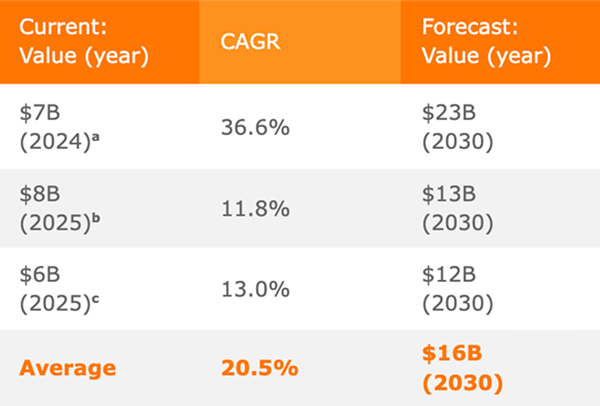
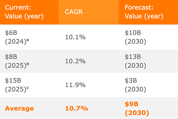
2.9 These next-generation antibodies generally are based on four development principles: improving the effector function and/or extending the half-lives of traditional mAbs, providing additional specificities for targeting, using smaller scaffolds, or conjugation to various small molecule actives.10 They are often more targeted and potent than traditional mAbs. The new approaches to designing multispecific drugs are also creating significant opportunities to develop new modes of action.11 Table 2 is a survey of different market forecasts for the growth of next-generation antibody therapeutics. Although the values vary, depending on the report, all of them predict this niche of the market is one of the fastest-growing aspects of the pharmaceutical industry.
3.1 While initial antibody therapeutics were derived from naturally-occurring antibodies, or antibodies developed against known targets, modern therapeutic development relies more heavily on developing novel antibodies against novel targets. A druggable target is typically identified during research into a particular disease, after which the researchers must go about designing (“discovering”) an antibody against it. Antibody discovery technologies can generally be categorized as in vitro or in vivo approaches. Each type of approach has distinct advantages and disadvantages, which are heavily debated in the literature and scientific meetings.
3.2 Historically, animals have been injected with the target antigen formulated in an appropriate adjuvant so that the host will develop an immune response. After a few weeks, the resulting B cells are isolated. The first techniques developed would require the recovered B cells to be hybridized with cancer cells so that the resulting hybridomas could be cultivated in the lab. The clonal hybridomas were then isolated and the product of each (a monoclonal antibody) assayed for the desired characteristics. More recently, primary B cells can be sorted by flow cytometry, assayed by ELISA methods, and/or sequenced without the need to generate hybridomas.
3.3 No matter the characterization process, antibodies derived from a nonhuman host must then be “humanized” so that the human immune system will not identify the protein as foreign. This was once done “by hand” with in silico design. In some cases, a common human antibody Fc sequence could simply be grafted onto the variable region sequences. Today, more advanced techniques are available. One method rapidly gaining acceptance is the creation of a transgenic animal with a humanized immune germline. These hosts will mount an immune response, but the resulting antibodies carry the human Fc sequence.
3.4 In vitro methods generally involve generation of a library of antibody sequences that are expressed on the surface of E. coli or yeast. The resulting library is screened against the antigen, and the resulting microbial clone is then isolated and characterized.
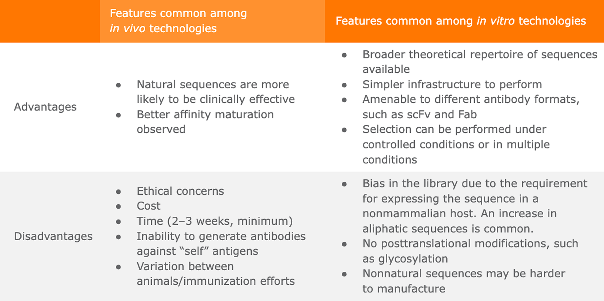
3.5 High-level advantages and disadvantages to each type of approach are summarized in Table 4.15 As technology advances, the relative disadvantages of each are becoming less prominent. For example, one major disadvantage of in vivo discovery is the need for an immunogenic target. Some companies are using hosts further removed genetically from humans for discovery, such as chickens or cows.16 The more distantly related hosts are more likely to generate a strong immune response to a human antigen, but again the antibodies generated need to be humanized. Humanization of antibody sequences has long been performed on mouse-derived (murine) antibodies and so the techniques for addressing this challenge are becoming more advanced.
3.6In vitro technologies enjoy a clear advantage in that they do not rely on an animal to mount an immune response. This broadens the range of targets that can be used and reduces the cost and ethical concerns of using animals. In vitro technologies, however, suffer from any bias in their libraries. Libraries are generally prone to bias based on expressibility in the host (yeast or E. coli), which, for example, tends to increase the prevalence of aliphatic sequences in the resulting clones. Nonnatural sequences can be difficult to manufacture or pose problems in the clinic. The resulting heavy and light chains may not be physically compatible, which decreases the stability of the antibody. Machine learning (ML) is being employed to create libraries and screen antibodies for affinity and developability by addressing each of these challenges. It is interesting to note that ML produces different results depending on whether the training data are derived from DNA sequencing or peptide sequencing.15 Clearly, innovation in the field of antibody discovery can be expected for some time to come.
3.7 As we have seen, global market forecasts vary among sources, but a short survey of forecasts for the antibody discovery market consistently predict healthy growth in this portion of the biopharmaceutical market (Table 3).
Table 4. Relative Advantages of In Vitro and In Vivo Technologies
4 Cell Line Development
4.1 A wide range of human and other mammalian cells are routinely cultured in the laboratory for research and diagnostic purposes, yet biologics production of glycosylated proteins such as antibodies is mostly confined to HEK and CHO cells. This is a result of a few different factors. Innovators, especially small biotechs, need to minimize the unknowns in their already risky development programs wherever possible and prefer to stick with cells that have a well-established set of protocols for culture, scale-up, and testing. There are a wide range of off-the-shelf resources to support the CHO and HEK cell–based programs, such as optimized feed media and residual DNA and host cell protein testing kits.
4.2 In addition to the practical reasons for using CHO and HEK, regulatory considerations are, of course, also a factor. The FDA requires extensive documentation of the source of any cells used in production so that the innovator can demonstrate a very low risk that some as-yet-undetectable adventitious agent is present in the cell bank. There are now many commercially available cell lines for which the cell line history data package has been approved many times by the FDA as a part of previous investigational new drug (IND) filings.
4.3 HEK cells are the host of choice for some clotting factors because the human glycosylation patterns are more critical for these long-term therapies. HEK cells have also found a resurgence with the rise of viral vector production, as the commonly used vectors grow better in HEK than CHO cells. For the most part, CHO cells are used for everything else that must be produced in mammalian cells. While the glycosylation patterns are not identical to human cells and vary widely depending on culture conditions, it has been sufficiently established that CHO glycosylation is compatible with human therapies. Innovators who are initiating a cell line development program are often bewildered by the cell line options and molecular tools available. In general, the host cell line is available for research use for a small fee. Here “research use” includes all development work up to the filing of an IND application.
4.4 Innovators typically need to pay a milestone fee upon IND filing, and additional milestones or royalty payments may be required as the program progresses through the clinic. Royalties are often required upon market approval. Thermo Fisher Scientific has one of the simpler license structures. Their Gibco Freedom CHO-S Kit cells are available as a catalog item for research use. When a customer wishes to file an IND, they pay a one-time fee to the company, upon which they gain access to the cell line data history package to support the IND documentation.20
4.5 Although the initial patents surrounding some key cell lines, notably the Lonza-GS system, have expired, the cost for licensing a cell line is still very high and terms are often murky until the innovator signs a confidentiality agreement. Vendors can command such high fees because customers need the reassurance of a track record with regulatory filings and practical support for the development programs.
4.6 The second generation of cell lines focuses more on the molecular aspect of cell line development. Vendors such as ATUM, ProteoNic, and Synthego have developed molecular tools that come with their own licensing fees, but these types of tools are largely responsible for the rapid increase in titers.21 Some often-overlooked aspects of cell line development improvements enabled by better molecular tools are greater stability and improved pool behavior.
4.7 The FDA’s Center for Biologics Evaluation and Research (CBER) states that the “growth pattern and morphological appearance of the cell line should be determined and should be stable from the master cell bank to the end-of-production cells.”22 Subsequent guidances recommend this can best be demonstrated by maintaining a continuous culture of the production cell through 60 generations. To achieve 60 generations, CHO cells must generally be maintained for six to eight weeks. When the stability of the top candidate clones is questionable, it may be prudent to wait for the stability data before committing to a clone and proceeding with the process development program and cell banking. If the stability of the top clones is nearly certain, it is likely to be worth the risk of continuing with the program while stability data is generated in parallel. This single characteristic of a host cell line can, therefore, save eight or more weeks in a program and is often highly valued by savvy innovators.
4.8 The COVID-19 pandemic accelerated the use and FDA acceptance of stable pools for production of phase I material.23 Because cloning can require eight weeks (before the additional eight weeks of stability testing) this is another point at which innovators can accelerate their programs. Molecular tools that create pools that behave consistently are essential for this approach. High titers are, of course, desired, but the consistent culture characteristics are critical.
4.9 Stability and consistent pool behavior often share the same molecular foundations and can usually be achieved through one of two mechanisms. The first is by limiting and controlling the locations at which the vector integrates into the chromosome to favor sites that are stable and maintain high expression. The second method is by insulating the vector sequence — no matter where it integrates in the chromosome — from any outside influence. The net effect is that expression from the vector in each cell within a pool behaves exactly as all of the other cells, even though the integrated plasmid is randomly located in a different spot on the chromosome.
4.10 As molecular tools become more prevalent, though still guarded by patents or trade secrets, we have seen a rise in innovators and CDMOs signing partnership licensing agreements with cell line development specialists to improve the titers and stability of their cell lines, often while shortening the overall timelines of their programs.
4.11 Once a production cell line is developed, selected, and cloned, it must be banked and characterized according to strict protocols meeting necessary compliance standards for potential future regulatory filings.24
4.12 Market numbers specific to standalone banking of production cell banks for biotherapeutics and vaccines are difficult to ascertain because of the larger market for generation and storage of stem cell banks and other applications, such as regenerative medicine, somatic cell therapy, gene therapy, and tissue engineering.
4.13 When we combine cell line development and cell banking sales forecasts even this relatively minor portion of the biologics manufacturing market is very large and predicted to grow at a rate similar to other services in the industry (Table 5).
Table 5. Market Forecasts: Cell Line Development and Cell Banking25–27
5 Leveraging Digital Technologies for mAb Design
5.1 As in many other aspects of biopharma discovery and development, digital technologies, most notably artificial intelligence (AI) and machine learning (ML), are playing a growing role in the discovery, design, and development of mAbs. Fully in silico mAb design leveraging AI and ML, to be truly effective, requires a deep understanding of the rules of mAb–antigen binding, the ability to combine different mAb design parameters in a modular fashion, and algorithms that enable unconstrained parameter-driven in silico mAb sequence synthesis. If done correctly, such in silico antibody discovery tools can achieve fast, inexpensive, and on-demand generation of fit-for-purpose antibodies.28
5.2 The application of digital technologies to antibody design has increased greatly in recent years. Several start-ups offer antibody design services, and many large biopharma companies have established their own internal expert teams with the intent of accelerating the development of more optimal antibodies from safety, efficacy, and manufacturability perspectives.
5.3 One recent example of an in silico approach to mAb development involved examination of the binding abilities of eight mAbs against several SARS-CoV-2 variants of alpha (B.1.1.7) and delta (B.1.617.2) lineages. Using the obtained data, a chimeric antibody was designed by conjugating the CDRH3 of regdanivimab with a sotrovimab framework to ensure that all variants are neutralized.29
5.4 In a separate example, using a semisynthetic design approach combining bioinformatics techniques and physical laboratory work, a new antibody library format was established that affords high-affinity binders with drug-like developability properties directly from initial selections, reducing the need for further engineering or affinity maturation.30 This approach should, according to the authors, reduce failure rates while also shortening development times.
6 In Vivo Antibody Production
6.1 The success of messenger RNA (mRNA) vaccines against COVID-19 has opened the door to many other potential mRNA-based vaccines and therapeutics. It has also drawn attention to the effectiveness of having the body produce the active drug or vaccine agent in vivo rather than via in vitro cell culture or bacterial/yeast fermentation. Researchers are consequently looking at ways to have healthy cells produce other drug substances, including antibodies.6
6.2 This in vivo approach is particularly attractive for the development of engineered antibody fragment-based therapeutics. These smaller antibody derivatives can achieve greater tumor penetration and enhanced epitope binding, especially for those epitopes not accessible by larger, whole antibodies. Antibody fragments have short half-lives, however, which has prevented their widespread development as therapeutics. A potential solution is to pursue antibody gene therapy, or the use of gene-based strategies for in vivo production of antibody fragments.
FAQS
What is the difference between IgG-like and non-IgG-like bispecific antibodies?
Answer: The primary distinction lies in the presence of the Fc (constant) region. IgG-like antibodies (e.g., CrossMab, DVD-Ig) include an Fc region, which grants them longer half-lives but makes them more difficult to manufacture. Non-IgG-like antibodies (e.g., BiTEs, DARTs) lack the Fc region; they are smaller, penetrate tumors more effectively, and are easier to produce, but they are cleared from the body much faster.
Why are CHO cells the industry standard for therapeutic antibody production?
Answer: CHO (Chinese Hamster Ovary) cells are preferred because they provide human-compatible glycosylation patterns and have a well-established regulatory track record. They support complex molecular tools like "knob-in-hole" structures that facilitate the correct assembly of multispecific antibodies, ensuring stability and reducing manufacturing risks for innovators.
How does AI improve monoclonal antibody (mAb) discovery?
Answer: AI and Machine Learning (ML) accelerate discovery by enabling unconstrained in silico sequence synthesis. These tools predict the "developability" and affinity of an antibody before physical lab work begins, addressing challenges like library bias and physical incompatibility between heavy and light chains, ultimately reducing failure rates and development timelines.
What are the clinical risks of bispecific T-cell engagers (BiTEs)?
Answer: The most significant clinical risk for bispecific antibodies, particularly T-cell engagers like blinatumomab, is Cytokine Release Syndrome (CRS). This exaggerated immune response is so common in this class that seven out of nine FDA-approved bispecifics carry a black box warning.
References:
Janeway, Charles A. Jr. et al. “The structure of a typical antibody molecule.” In: Immunobiology: The Immune System in Health and Disease. 5th edition. New York: Garland Science, 2001.
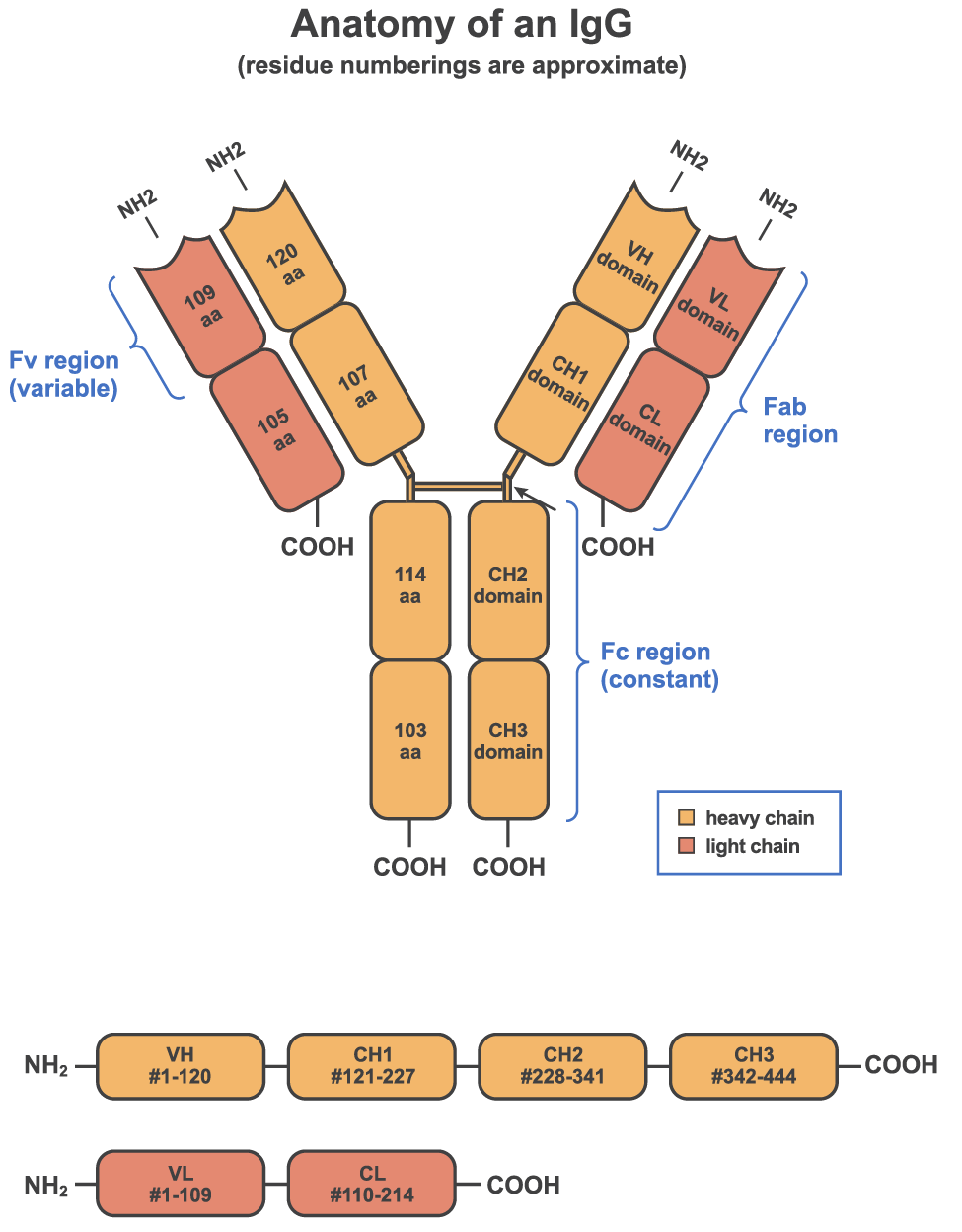 Figure 1. Structure of an Antibody
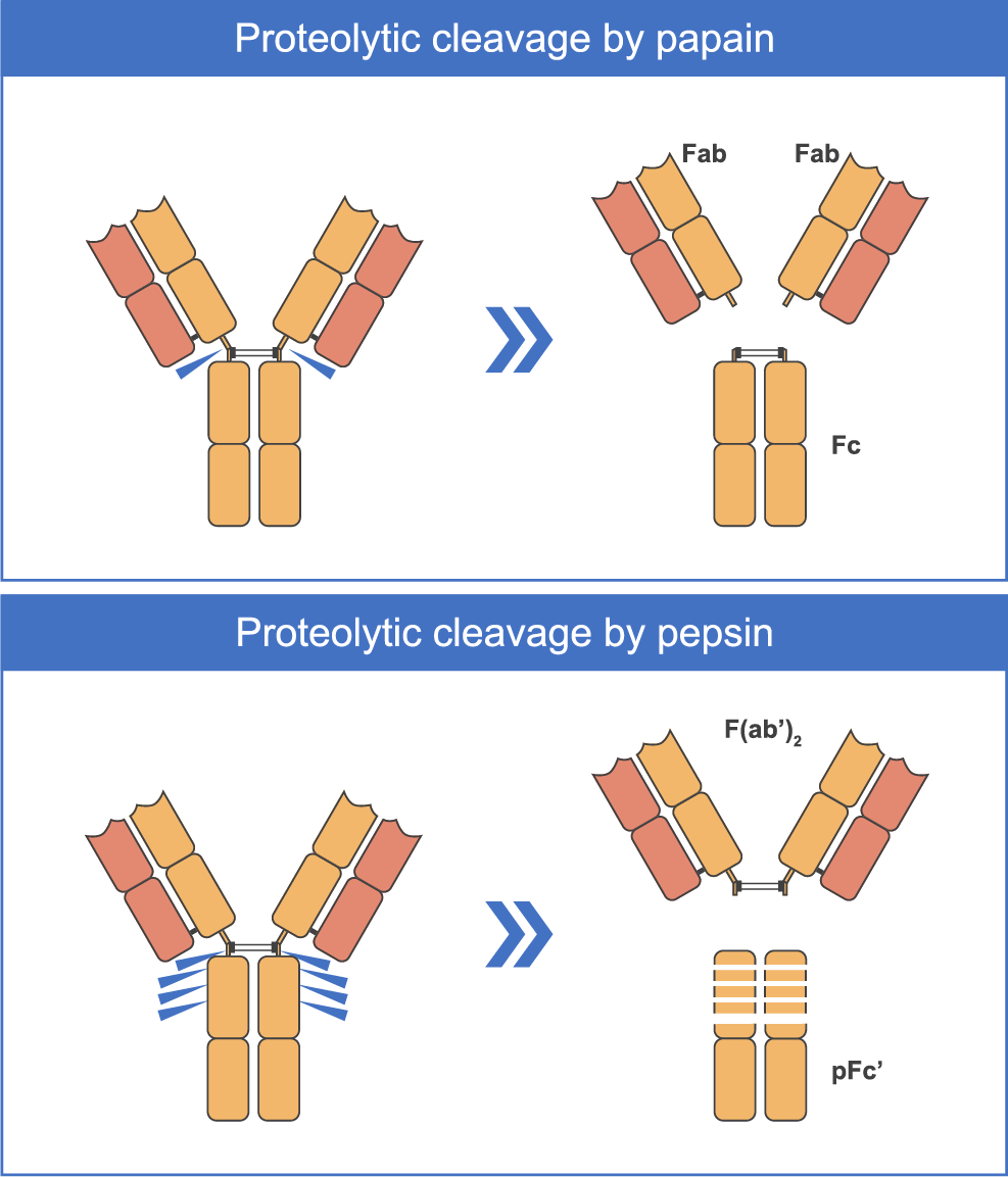 Figure 2. Common Antibody Fragments Created by Protease Cleavage
 2.2 The larger formats that contain Fc regions are generally more difficult to manufacture and are almost always produced in Chinese hamster ovary (CHO) cells. To facilitate assembly, a knob-hole structure is often incorporated into the CH domains. This aids assembly by reducing the affinity between the two like pairs and enhances binding between the different heavy chains. Since the approval of blinatumomab in 2014, eight additional bispecific antibodies have been approved by the FDA. Of the nine total approved bispecific drugs, all but two (amivantamab-vmjw and faricimab-svoa) have a black box warning due to the risk of cytokine release syndrome (CRS).
 3 Survey of Discovery Technologies
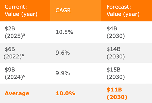 Table 4. Relative Advantages of In Vitro and In Vivo Technologies
 4 Cell Line Development
 5 Leveraging Digital Technologies for mAb Design